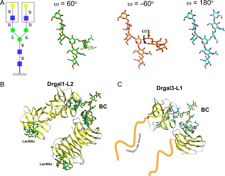
Fig. 3.
Model of the biantennary carbohydrate. (A) Common biantennary N-glycan observed in IHNV and fish epithelia. Monosaccharide symbols follow the symbol nomenclature for glycans system (PMID 26543186, Glycobiology 25:1323–1324, 2015). The three lowest energy conformations of the carbohydrate as calculated by the Glycam server (http://glycam.org). (B) Recognition of BC by two Drgal1-L2 dimers (left) and two Drgal3-L1 CRDs (right).

