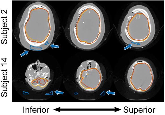
Fig. 8.
Example cortical surface segmentation results using a deep convolutional neural network (DCNN) where predictions were poor. Segmentation contours show (i) the initial DCNN segmentation (blue contour), (ii) the segmentation result after post-processing (orange contour), and (iii) the ground-truth segmentation (yellow contour). Axial slices are shown for each subject progressing from the bottom of the head (left) up to the top (right). Arrows highlight areas where the DCNN produced incorrect segmentation results.

