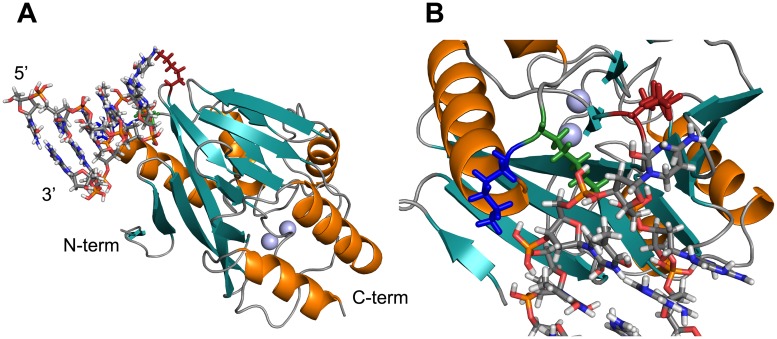
Fig 5. Model of the 10-mer bound to 5/B/6 MBL provides a picture of the inhibited state.
Model of the 10-mer bound to 5/B/6 MBL. The lowest energy HADDOCK structure is given, while a representative structure from the seven other clusters is given in S5 Fig. The coloring of secondary structural elements for the 5/B/6 MBL follows Fig 2, and the red, blue and green sticks denote K78, K104, and K107, respectively. The 10-mer is shown as salmon sticks and the two Zn2+ ions are light blue spheres. A) The overall structure of the enzyme-10-mer complex. B) A closer view of the enzyme-10-mer interaction site.

