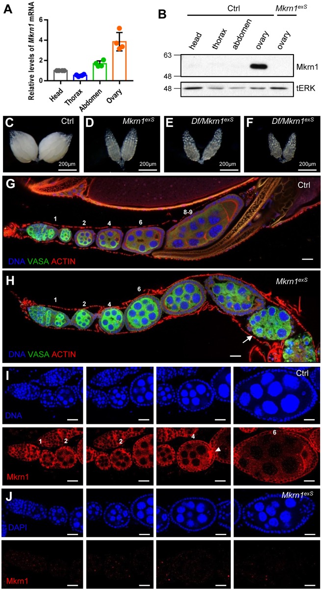
Fig 1. Mkrn1 expression was highly enriched in control ovaries and Mkrn1-null ovaries exhibited a phenotype.
(A) One-day-old, control female flies from standard nutrient conditions were dissected for baseline analyses. Total mRNA was extracted from the heads, thoraxes, abdomens, and ovaries and analyzed by quantitative real-time PCR. Relative mRNA levels for Mkrn1 and reference gene Actin are shown. Error bars represent the SEM from four independent experiments. (B) Protein extracts from indicated tissues were analyzed by immunoblot using the anti-Mkrn1 antibody and tERK was used as a loading control. (C–F) Bright-field images of whole ovaries from control (C), Mkrn1exS (D), Df(3L) BSC418/Mkrn1exS (E), and Df(3L) BSC419/Mkrn1exS flies. (G, H) Confocal images of ovariole immunostaining from control (G) and Mkrn1exS (H) flies. Dissected ovaries were stained for Vasa (green), Actin (red, phalloidin), and the DNA (blue, hoechst). Please note that Mkrn1exS egg chambers did not progress to vitellogenic stages and eventually degenerated from stage 7 resulting in egg chambers with only germline cells (arrow). (I, J) Ovaries were immunostained for DNA (blue, top panel) and Mkrn1 (red, bottom panel) from control (I) and Mkrn1exS (J) flies. Mkrn1 was ubiquitously observed in cytoplasm of follicle cells, nurse cells, and oocytes throughout oogenesis, and Mkrn1 was enriched in the pole plasm (arrow head) in the control. Absence of Mkrn1 detection in Mkrn1exS ovaries validates the gene knockout and specificity of the Mkrn1 antibody. Numbers above each egg chamber indicate the developing stage of ovarioles. Scale bars = 20μm.

