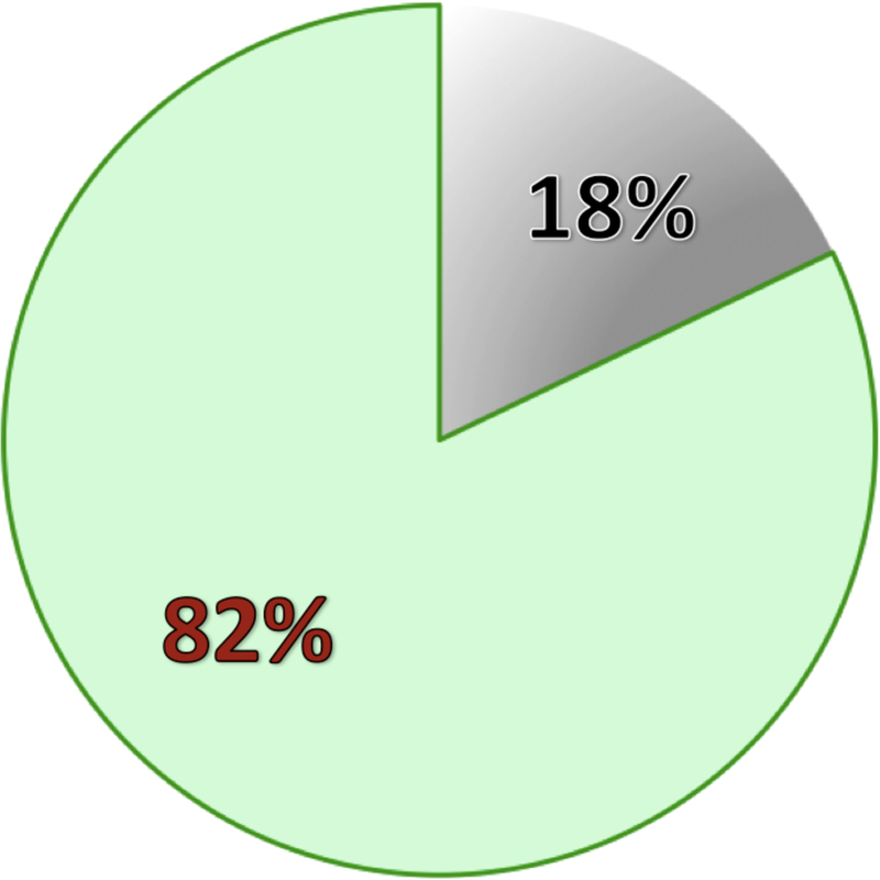
Figure 4: Ratio of electrodes in epileptic and non- epileptic tissue.

In 100 patients implanted with ECoG or sEEG electrodes at Stanford Medical Center, we reviewed the iEEGs in each patient and labeled pathological electrodes that contained epileptic activity (i.e., recorded seizures or epileptiform spikes). We used the total number of electrodes implanted in these patients to calculate the ratio of pathological (gray) to non-pathological (green) sites. In patients with focal epilepsy, sites with pathological activity are clustered to few electrode contacts. The extent of non-pathological electrodes will depend on the density, form, and size of implanted intracranial electrodes. In a patient with wide coverage and focal epileptic zone, non-pathological sites will be covered across a large region of the brain.
