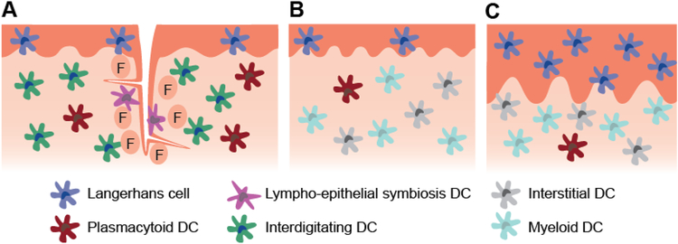
Figure 1.
Schematic representation of the mucosal structure and select dendritic cell subsets present in the a) tonsils of the WTR, b) sublingual mucosa, c) buccal mucosa. The lingual and palatine tonsils are covered with ~100 μm thick stratified squamous epithelium and contain crypts lined by a non-uniform lymphoepithelium. The outer epithelium primarily contains Langerhans cells, while the crypt epithelium contains lympho-epithelial symbiosis DCs along with macrophages and lymphocytes (not depicted). Other DC populations including plasmacytoid DCs and interdigitating DCs densely populate the follicular region (F). Germinal center DCs are also present within germinal centers (not depicted). The sublingual epithelium ranges from 100–200 μm thick, while the buccal epithelium ranges from 500–800 μm thick. The buccal epithelium contains approximately 2.5 times the number of Langerhans cells in the sublingual epithelium. Submucosal DC subsets are not well characterized in humans, but evidence suggests that interstitial and myeloid DCs are present and plasmacytoid DCs may infiltrate during inflammation.

