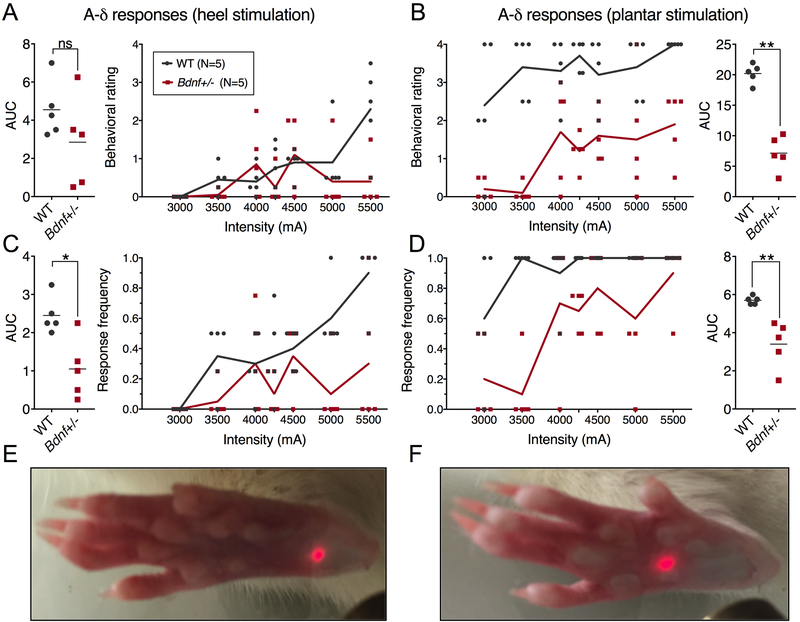
Figure 4. Short-pulse A-δ laser stimulation of WT and Bdnf+/− heel and plantar surface of the hind paw.
A 100ms pulse was delivered to the heel (A, C, E) or plantar surface (B, D, F) across a range of stimulus intensities, and behavioral assessments were scored by a blinded observer (N=5). Ratings of behavioral responses were made using a previously validated scale [41; 42], with significant differences observed on the plantar (B), but not the heel (A). The plantar area is more sensitive to thermal stimulation than the heel, resulting in lower overall responsiveness to the same stimulus intensity on the heel (C,D) Response ratios to hind paw stimulation were significantly different (P<0.01)for both the heel and plantar stimulation paradigms. Differences in average area under the curve (AUC) measurements were calculated using a Wilcoxon Mann-Whitney U-test; *, p < 0.05; **, p < 0.01.

