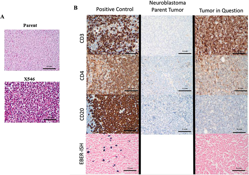
Figure 1.
A. Hematoxylin and eosin staining of the PDX tumor in question revealed a tumor composed of small blue round cells (bottom panel), not significantly different from the parent (top panel). B. Side by side comparison of the PDX in question (X546), a known human neuroblastoma, and positive control demonstrated marked differences in staining between the transformed PDX and the parent tumor. The PDX in question (right panels) stains positive for CD3, about 5% positive for CD4, and negative for CD20, while the patent tumor (middle panels) stains negative for all three immunomarkers. EBER-ISH for the PDX was negative (bottom right panel) compared to positive control (bottom left panel), indicating that this lymphoma was not EBV related.

