Figure 1.
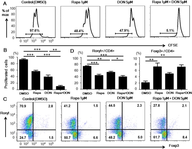
Rapamycin with DON additively suppressed CD4+ T cell proliferation in vitro. (A,B) CD4+ T cells, labeled with carboxyfluorescein diacetate succinimidyl ester (CFSE), were isolated from untreated Balb/c mice, placed in culture plates precoated with anti-CD3 and CD28 mAbs, and cultured with either DMSO (control), 1 μM rapamycin (Rapa), 5 μM DON, or the combination of 1 μM rapamycin and 5 μM DON (Rapa + DON). After 3 days of culture, the frequency of proliferating CD4+ T cells was assessed by measuring the CFSE fluorescence using flow cytometry. Data are representative of 4 independent experiments. (C,D) CD4+ T cells isolated from untreated Balb/c mice were placed in culture dishes precoated with anti-CD3 and anti-CD28 mAbs, and cultured in medium containing IL-17, TGF-β, anti-IFN-γ, anti-IL-4 antibodies, and one of the four drug regimens described in (A). The cells were collected on day 5 of culture and stained with anti-CD4, anti-Rorγt, and anti-Foxp3 antibodies, and the frequencies of Th17 (Rorγt+/CD4+) and Treg (Foxp3+/CD4+) cells were analyzed by flow cytometry. Data are representative of 4 independent experiments. Bars show mean ± SEM. *P < 0.05; **P < 0.01; ***P < 0.001.

