
FIGURE 2.
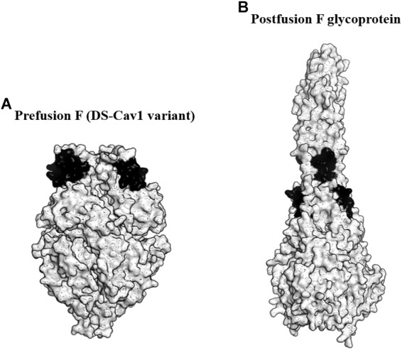
Targeting the prefusion F glycoprotein of respiratory syncytial virus (RSV). (A) Surface representation of the DS-Cav1 F glycoprotein variant (PDB ID: 4MMU). DS-Cav1 adopted the prefusion F glycoprotein conformation. The antigenic site ø is shown in black. (B) Surface representation of the trimeric postfusion F glycoprotein (PDB ID: 3RKI). The disrupted antigenic site ø caused by structural rearrangement from the prefusion to postfusion F is shown in black.