
Figure 1.
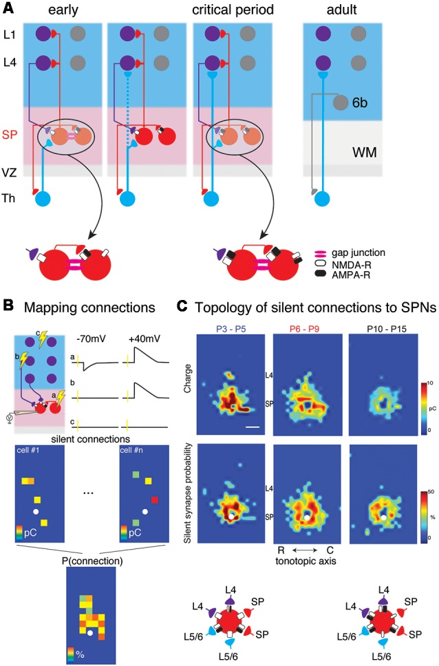
(A) Changing circuits between thalamus (Th), subplate (SP) and cortical layers 4 (L4) and 1 (L1). Insets show the existence of NMDA-R only “silent” synapses at early ages. (B) Cartoon illustration mapping of connections to subplate neurons (SPNs) via laser-scanning photostimulation (LSPS). Patch clamp recordings in voltage clamp are made from SPNs and locations are selectively stimulated. The SPN is recorded at −70 mV and +40 mV membrane potential at each stimulation location. Traces (a–c) indicate three potential outcomes of stimulation at the respective locations. (a) Stimulation of presynaptic locations that were connected to the SPN with AMPA and NMDARs resulted in excitatory postsynaptic currents (EPSCs) at both −70 mV and +40 mV. (b) Stimulation of presynaptic locations that were connected to the SPN with only NMDARs resulted in EPSCs at only +40 mV. (c) Stimulation of presynaptic locations that were not connected to the SPN resulted in no EPSCs at either −70 mV and +40 mV. Bottom shows calculating the spatial connection probability from maps of NMDAR-only connections (e.g., sites similar to site b). White circle indicates soma location of SPN. Maps of individual neurons are aligned to the soma and the probability of observing an EPSC is calculated for all relative spatial locations. (C) Changing topology of silent synapses over development. Shown are average connection probability and mean charge maps from LSPS experiments (from Meng et al., 2014). Cartoon below summarizes these data. At early ages, silent synapses are present between L4, L6/6, SP and SP and have a large synaptic strength. Silent synapses are most abundant at P6-P9 and at older ages, the strength of silent synapses has decreased.