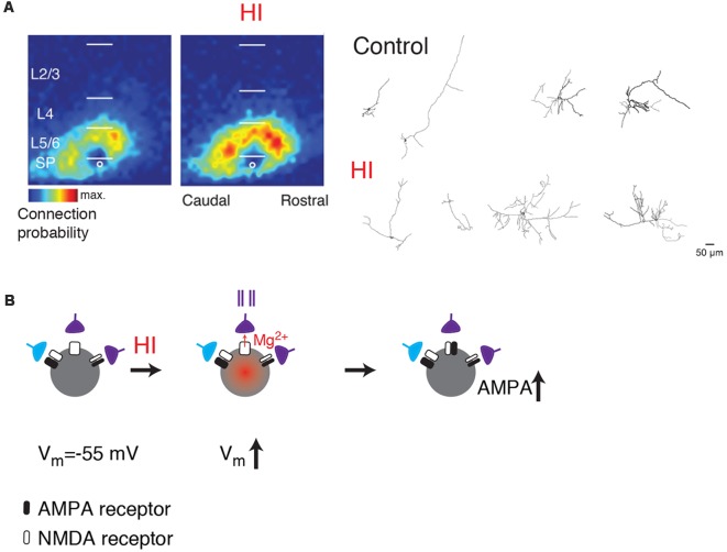
Figure 3.
(A) SPNs show functional and morphological hyperconnectivity after hypoxic-ischemic (HI; from Sheikh et al., 2019; no copyright permission as required for use of this image). Shown are LSPS maps of connection probability and neurolucida reconstructions. (B) Cartoon illustrating how depolarization caused by HI can lead to unsilencing of synapses on SPNs (middle). If presynaptic cells are active (indicated by spike train), the coincidence between presynaptic activity and postsynaptic depolarization might result in a subsequent increase in AMPARs (right, black).

