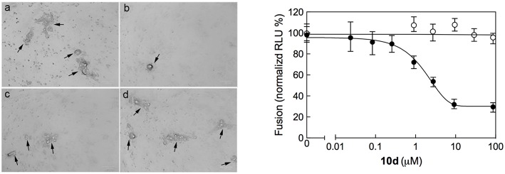
Figure 3.
(Left) Inhibition of RSV A2-induced syncytium formation in Vero-76 cells by 10d at a concentration of 0 μM (a), 50 μM (b), 10 μM (c), and 2 μM (d). Inoculum was removed 2 h post infection (p.i.), cells were left untreated or incubated 8 h p.i. with 10d, and syncytia formation was assessed 72 h p.i. as described in Materials and Methods. In the presence of 10d both size and number of syncytia did not increase. Images were taken 72 h p.i. using the ZOE Fluorescent cell imager (Bio-Rad) (bar size = 100 μm). Syncytia are indicated by black arrows. (Right) Quantitative dose-response cell-to-cell fusion assay using the DSP-chimeric reporter proteins and the ViviRen renilla luciferase substrate in the presence of compound 10d (filled symbols). The MeV (Measles Virus) F and H glycoprotein expression constructs (open symbols) were included for selectivity control. Reported values are normalized for DMSO-treated samples and are expressed as the mean of three experiments ± standard deviation. The EC50 value was obtained by 4-parameter variable slope regression fitting.

