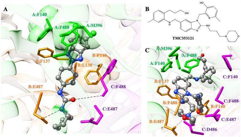
Figure 5.
(A) Putative binding mode of compound 10d into the 3-fold symmetric trimeric RSV F-protein in its pre-fusion state. The compound is shown as atom-colored balls-and-sticks (gray, C; blue, N; red, O; green, Cl). The three F protomers are represented as colored ribbons (light green, protomer A; light orange, protomer B; light purple, protomer C). The protein residues mainly involved in 10d binding are evidenced and labeled. Hydrogen bonds are depicted as broken black lines. Hydrogen atoms, water molecules, ions and counterions are omitted for clarity. (B) Structure of the known RSV F-protein inhibitor TMC353121. (C) Details of TMC353121 in the binding pocket of the three-fold symmetric trimeric RSV F-protein in its pre-fusion state. Representations and colors as in (A).

