FIGURE 2.

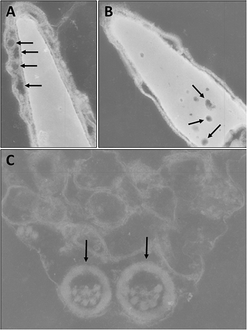
Transmission electron microscopy of sperm. (A) Abnormal spermatozoon with acrosomal vacuolation (arrows). (B) Abnormal spermatozoon with nuclear vacuolation (arrows). (C) Abnormal axoneme formation (arrows).

Transmission electron microscopy of sperm. (A) Abnormal spermatozoon with acrosomal vacuolation (arrows). (B) Abnormal spermatozoon with nuclear vacuolation (arrows). (C) Abnormal axoneme formation (arrows).