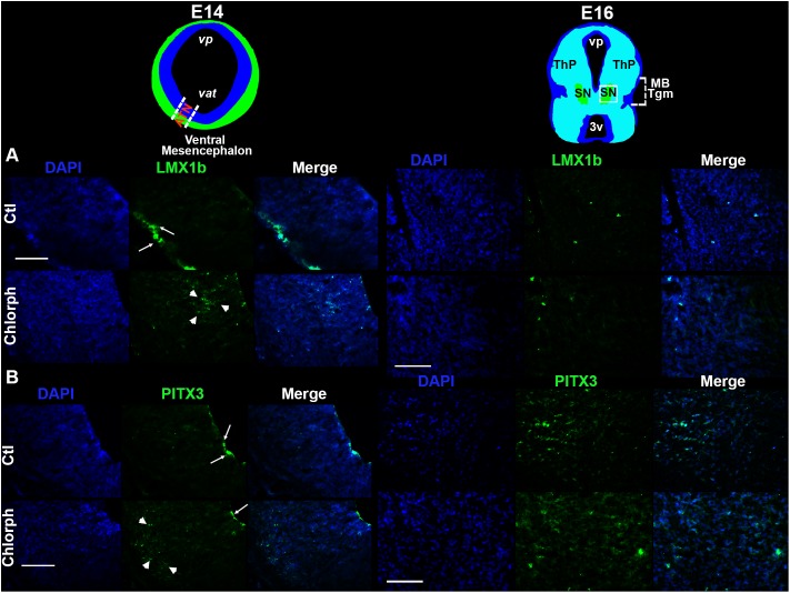
FIGURE 3.
Effect of chlorpheniramine administration to pregnant rats on dopaminergic commitment and differentiation in the ventral mesencephalon of E14 and E16 embryos. Top panel, coronal views of the mesencephalon at E14 (in blue is the ventricular zone and in green the marginal zone or differentiated field) and E16 (substantia nigra in green). The outlined area in white correspond to the area where epifluorescence micrographs (20×) were taken. (A,B) Representative micrographs showing individual and merged channels for LMX1b immunoreactivity (green in A; arrows shown the mark in the marginal zone and arrow heads in the subventricular zone of the mesencephalic neuroepithelium) and PITX3 (green in B; arrows show the mark in the ventricular zone and arrow heads in the differentiation field of the mesencephalic neuroepithelium), and nuclei stained with DAPI in blue in coronal sections of E14 and E16 embryos from control (Ctl) and chlorpheniramine-treated (Chlorph) rats. ATN, anterior tegmental neuroepithelium; vp, aqueduct pretectal; vat, aqueduct tegmental; MB Tgm, Midbrain Tegmentum; SN, substantia nigra; ThP, thalamus posterior; 3v, third ventricle. Scale bar, 50 μm.

