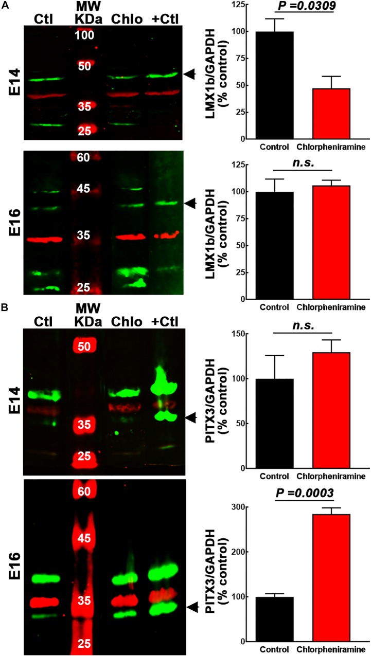
FIGURE 4.
Quantitative analysis of the effect of chlorpheniramine administration to pregnant rats on dopaminergic commitment and differentiation in the ventral mesencephalon of 14- and 16-day-old embryos. Left columns show representative Western blots for LMX1b (green, 41 kDa) in panel (A) and PITX3 (green, 32 kDa) in panel (B), for E14 and E16 ventral mesencephalon (vMes) from embryos from control (Ctl) and chlorpheniramine-treated (Chlo) rats. The internal control (GAPDH, 37 kDa) appears in red. Protein extracts from vMes E12 or adult substantia nigra were used as positive controls (+Ctl) for LMX1b and PITX3, respectively. MW, molecular weight ladder in kDa. Right columns, semi-quantitative fluorometry analysis of LMX1b (A) and PITX3 (B), for E14 and E16 tissues from the vMes. Values are expressed as percentage of the fluorescence ratio of the control tissues and are means ± SEM from four experiments. P-values were obtained after two-tailed unpaired Student’s t-test; n.s., non-significant.

