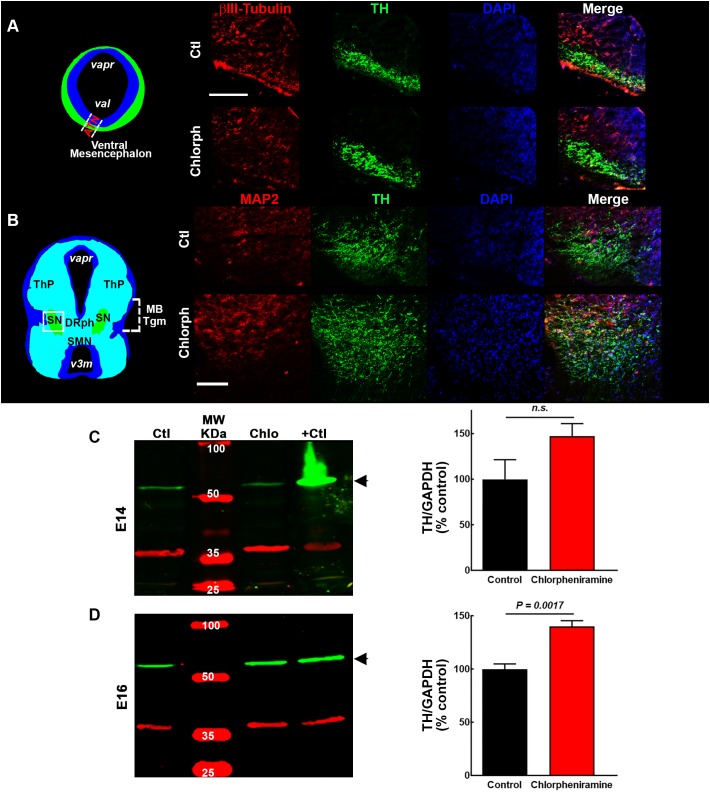
FIGURE 5.
Effect of chlorpheniramine administration to pregnant rats on tyrosine hydroxylase (TH) immunoreactivity and protein levels in the ventral mesencephalon of 14- and 16-day-old embryos. (A,B) Left, coronal view of the mesencephalon at E14 and E16, respectively. The area outlined in white dashed lines and square corresponds to the area shown in the representative epifluorescence micrographs (20×) on the right, which show independent and merged channels for βIII-tubulin (A) and MAP2 (B) in red, TH in green, and DAPI (blue) for the vMes from embryos from control (Ctl) and chlorpheniramine-treated (Chlorph) rats. ATM, anterior tegmental neuroepithelium; varp, aqueduct pretectal; vat, aqueduct tegmental, MBTgm, Midbrain Tegmentum; SN, substantia nigra; ThP, thalamus posterior; DRph, dorsal raphe; SMN, supramammillary nucleus; v3m, third ventricle. Scale bar, 50 μm. (C,D) Left columns, representative Western blots for TH (green, 60 kDa) and the internal control GAPDH (red, 37 kDa), for the vMes from embryos from control (Ctl) and chlorpheniramine-treated (Chlo) rats. Protein extracts from adult substantia nigra were used as positive controls (+Ctl). MW, molecular weight ladder (kDa). Right column, quantitative fluorometry analysis of TH at E14 (C) and E16 (D). Values are expressed as percentage of the fluorescence ratio of the control tissues and are means ± SEM from four experiments. P-values were obtained after two-tailed unpaired Student’s t-test; n.s., non-significant.

