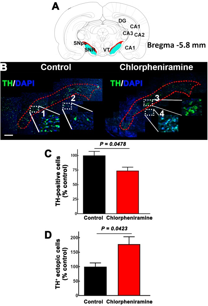
FIGURE 7.

Effect of chlorpheniramine administration to pregnant rats on the number of tyrosine hydroxylase positive cells in the substantia nigra pars compacta of P21 pups. (A) Cartoon of a coronal section from Bregma –5.8 mm in red the substantia nigra pars compacta. Modify from Paxinos and Watson. (B) Merged representative epifluorescence micrographs (20×) for TH immunoreactivity (green) and DAPI (blue, nuclei) in the substantia nigra pars compacta (outlined in red dotted lines) of coronal brain sections (Bregma –5.8 mm) from pups from control and chlorpheniramine-treated rats. The white dotted rectangles represent digitalized zoomed areas (3.5×) from control (1, substantia nigra and 2, ectopic mark), and chlorpheniramine (3, substantia nigra and 4, ectopic mark) TH+ cells. Scale bar, 200 μm. (C,D) Quantification of TH+ cells. Values are expressed as percentage of the mean of total TH+ cells of the control, and are means ± SEM from three experiments (three consecutive sections were used per experiment). P-values were obtained after two-tailed unpaired Student’s t-test. SNpc, substantia nigra pars compacta; VT, Ventral tegmental area; SNR, substantia nigra reticulata; DG, dentate gyrus; Cornu Ammonis areas, CA1, CA2, and CA3.
