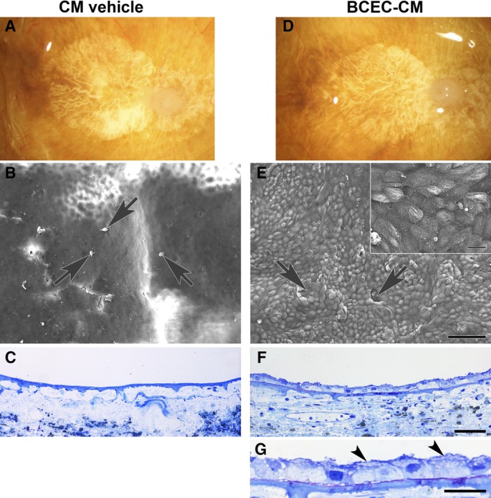
Figure 2.

Paired explants from an 82‐year‐old woman with geographic atrophy, seeded with fetal retinal pigment epithelium (RPE) cells. The patient's clinical history noted age‐related macular degeneration (AMD) for 20 years. (A, D): Post‐mortem clinical photographs showing subfoveal geographic atrophy before RPE cell seeding. In conditioned media (CM) vehicle, (B) only a few dead cells (arrows) and cellular debris are present on the explant surface. (C): No cells are present on Bruch's membrane surface. In bovine corneal endothelial cell (BCEC)‐CM, (E) RPE cells fully resurface Bruch's membrane in the area of geographic atrophy with a few very small defects (arrows). Localized areas of multilayering are present. Cell surfaces show abundant apical processes (inset). (F): In this field, cells resurfacing the BCEC‐CM explant are predominantly bilayered. Cells directly on Bruch's membrane are small and tightly packed; flat cells appear to overlie the cells in contact with Bruch's membrane. (G): Flattened cell processes overlying cells on top of Bruch's membrane are indicated by arrowheads. The cell processes contain vesicles. CM vehicle nuclear density (ND), 0; BCEC‐CM ND, 19.61 ± 0.43. Scale bars: 100 μm (E); 20 μm (E, inset); 50 μm (F); 20 μm (G). Toluidine blue staining. Reproduced with permission from Sugino et al. 58.
