Figure 1.

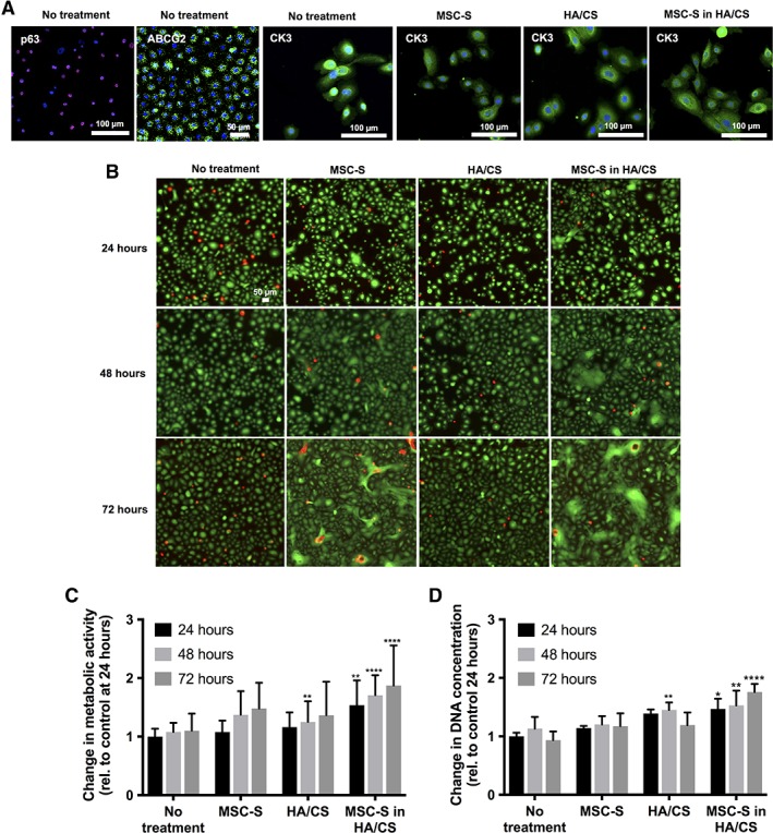
(A): P63, ABCG2, and CK3 staining were observed in the harvested primary human corneal epithelial cells (HCECs) used in this study, indicating a mixed population of limbal and central stem cells. CK3 was expressed in these cells 24 hours after treating with complete keratinocyte serum‐free medium (KSFM) with growth factors (no treatment, control), MSC‐S, HA/CS, MSC‐S in HA/CS. (B): Live/dead cytotoxicity assay after treatment with complete KSFM with growth factors, MSC‐S, HA/CS, and MSC‐S in HA/CS over 72 hours in primary corneal epithelial cells. (C): Effect of MSC‐S, HA/CS, and MSC‐S in HA/CS on primary HCECs proliferation. Cell proliferation was determined using a cell metabolic activity assay, and (D): DNA concentration, at 24, 48, and 72 hours. The data were normalized to no treatment at 24 hours. In both assays, proliferation was statistically significantly greater when MSC‐S was delivered in HA/CS compared to no treatment (n = 4; *, p < .05; **, p < .01; ****, p < .0001). Abbreviations: CS, chondroitin sulfate; HA, hyaluronic acid; MSC‐S, mesenchymal stem cells secretome.
