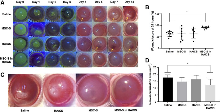
Figure 4.

Results from corneal alkaline burn model experiments in rats in vivo. Sodium hydroxide (NaOH) was applied to the epithelium followed by copious irrigation with balanced salt solution, followed by treatments (saline, MSC‐S, HA/CS, MSC‐S in HA/CS) 1 drop daily for 7 days and the corneas were photographed with and without fluorescein staining out to day 14. (A): Representative photographs for each of the study arms. Yellow arrows show areas of scar formation in the saline group and intrastromal hemorrhage in the HA/CS group (B): Percent (%) wound closure 24 hours after treatment. Eyes treated with MSC‐S in HA/CS yielded significantly smaller wound areas compared to the saline group (*, p < .05), while the HA/CS and MSC‐S treatments alone did not. (C): High‐magnification photographs of representative photos of alkaline burned corneas 14 days after saline, HA/CS, MSC‐S, MSC‐S in HA/CS. (D): Area of corneal neovascularization (NV) 14 days after treatments. The corneas treated with MSC‐S in HA/CS resulted in a lower NV area compared to the saline group (*, p < .05), whereas the HA/CS and MSC‐S groups did not (representative photographs taken at 15× magnification). Abbreviations: CS, chondroitin sulfate; HA, hyaluronic acid; MSC‐S, mesenchymal stem cells secretome.
