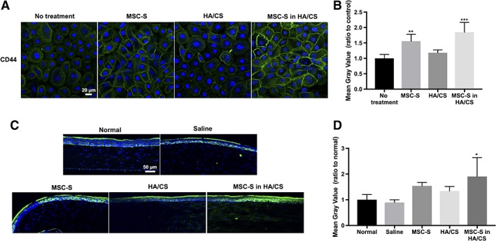
Figure 6.

(A): CD44 expression (green fluorescence) in human corneal epithelial cells after treatment with complete keratinocyte serum free medium with growth factors (no treatment, control), MSC‐S, HA/CS, MSC‐S in HA/CS—scale bar 20 μm. (B): Quantification of the fluorescence intensities using ImageJ revealed that CD44 receptors are upregulated in the cells that received MSC‐S (**, p < .01) and MSC‐S in HA/CS (***, p < .001). (C): CD44 expression in rat corneas in vivo 7 days after treatment with saline, MSC‐S, HA/CS, MSC‐S in HA/CS, and uninjured/untreated corneas—scale bar 50 μm. (D): Quantification of the fluorescence intensities using ImageJ showed that CD44 receptors are upregulated in the cornea treated with MSC‐S in HA/CS (*, p < .05). Abbreviations: CS, chondroitin sulfate; HA, hyaluronic acid; MSC‐S, mesenchymal stem cells secretome.
