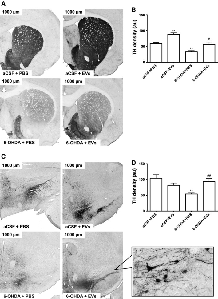
Figure 6.

Influence of intranasally administered extracellular vesicles on expression of tyrosine hydroxylase in the substantia nigra (SN) and striatum of 6‐OHDA‐treated rats. Representative photographs show rat striatum (A) and SN (C) at ×300 magnification. Density measurements (a.u.) are shown in (B) for the striatum and in (D) for the SN. Inset of (C) depicts SN at higher magnification (×600). Values are expressed as mean ± SEM one‐way ANOVA followed by Tukey's post‐test. *, p ≤ .05 versus aCSF+PBS; # , p ≤ .05 versus 6‐OHDA+PBS.
