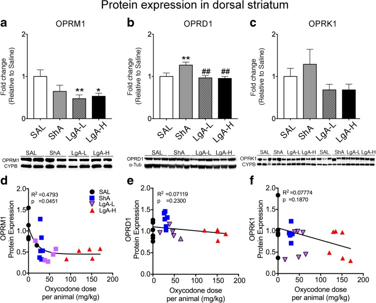
Fig. 3.
Mu receptor protein levels are decreased in the dorsal striatum of LgA rats after a month of forced abstinence. a–c Quantification of protein expression and representative images of Western blots showing levels of mu (OPRM1), delta (OPRD1), and kappa (OPRK1) proteins in rat striata. a LgA-L and LgA-H show decreased striatal OPRM1 protein levels. b Striatal OPRD1 protein levels show significant increases in the ShA rats. c Striatal OPRK1 protein levels were not significantly affected in any of the groups. d OPRM1 protein expression shows negative correlation to individual oxycodone intake. e OPRD1 and f OPRK1 protein expression show no significant relationship to drug intake. For quantitative Western blot analysis, the bands were normalized to cyclophilin B (CYPB) or α-tubulin (α-Tub). The values in the bar graphs represent means ± SEM (n = 5–6 rats per group). Key to statistics: *, **, p < 0.05, 0.01, respectively, in comparison to saline rats; ##p < 0.05 in comparison to ShA rats

