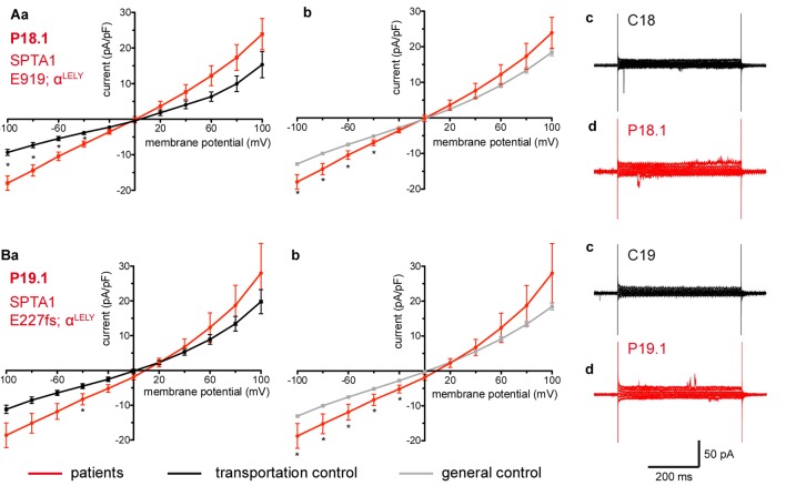
FIGURE 3.
Whole-cell recordings of ion currents from RBCs of healthy donors and HS patients with α-spectrin mutations and carrying at the same time an αLELY allele. Compared are the I/V curves of P18.1 (n = 10) with its own transportation control C18 (n = 6) (Aa) as well as with a general control (n = 175) (Ab), where n denotes the number of cells from the patient or the controls. As examples raw current traces recorded from the RBCs of a healthy donor (C18), whose blood was delivered together with the blood of P18.1 (Ac) and of patient P18.1 (Ad) are presented. Capacitances were not any different either with the transportation control (0.59 pA/pF P18.1 vs. 0.74 pA/pF C18; p > 0.05) or with the general control (0.59 pA/pF patient vs. 0.69 pA/pF control; p > 0.05). Compared are the I/V curves of P19.1 (n = 6) with its own transportation control C19 (n = 7) (Ba) as well as with a general control (n = 175) (Bb), where n denotes the number of cells from the patient or the controls. As examples raw current traces recorded from the RBCs of a healthy donor (C19), whose blood was delivered together with the blood of P19.1 (Bc) and of patient P19.1 (Bd) are presented. Capacitances were not any different either with the transportation control (0.66 pA/pF P19.1 vs. 0.58 pA/pF C19; p > 0.05) or with the general control (0.66 pA/pF patient vs. 0.69 pA/pF general control; p > 0.05). Significant differences are determined based on an unpaired t-test with ∗ representing p < 0.05. Mutations below patients numbers are designated as amino acid substitutions in the respective protein. The label αLELY next to the mutation stands for the presence of an αLELY allele in the corresponding patient.

