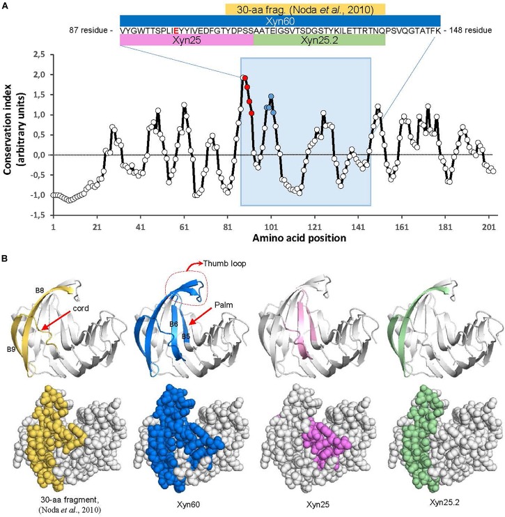
FIGURE 1.
Location of BcXyn11A peptides in the protein sequence and 3D model. (A) Evolutionary conservation of the whole BcXyn11A mature sequence calculated with AL2CO (http://prodata.swmed.edu/al2co) for an alignment of 1469 GH11 xylanases downloaded from Pfam (https://pfam.xfam.org) and aligned with Clustal Omega (https://www.ebi.ac.uk/services). Conservation indexes were averaged in windows of five residues to smooth the graph. The position of the Xyn60 peptide is indicated with a blue background. Inset shows the corresponding sequence to Xyn60 (residues 88 to 147 of BcXyn11A sequence expressed in P. pastoris, Supplementary Figure S1) and the relative position along the BcXyn11A sequence of the peptides used in this study. One of the two Glu residues (E98) involved in catalysis (Noda et al., 2010) is shown in red. The position of the M1and M2 regions (see below) are pointed as red and blue points, respectively. (B) Location of the peptides in a 3D model of BcXyn11A obtained in silico (Biasini et al., 2014). Notable structural elements, the thumb, the cord and the palm (Paes et al., 2012) are indicated.

