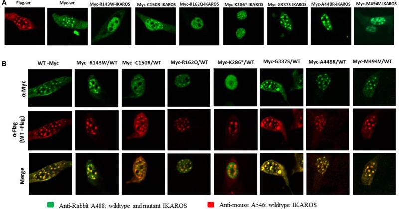
Figure 4.
The results of confocal microscopy with epitope-tagged constructs were consistent with the results of Immunofluorescent microscopy. (A) NIH3T3 cells, transiently overexpressing epitope-myc/FLAG-tagged wildtype IKAROS and myc-tagged mutant forms were incubated with monoclonal rabbit anti myc-tag-antibody and mouse monoclonal anti-FLAG M2 antibody and stained with goat anti-rabbit Alexa fluor 488 (green) and goat anti-mouse Alexa 546 (red). In NIH3T3 cells transfected with epitope-myc/FLAG-tagged wildtype IKAROS the speckled nuclear localization was observed in contrast to a diffuse nuclear staining, observed in NIH3T3 cells expressing the myc-tagged Arg143Trp, Cys150Arg, Arg162Gln, and Lys286* mutant forms. (B) Wildtype components were dominant to form IKAROS complexes in PC-HC sites. NIH3T3 cells were co-transfected with 50% of myc/FLAG-tagged wildtype IKAROS and 50% of myc-tagged, expressing each mutant form. The diffuse nuclear localization of the mutant forms was significantly reduced in NIH3T3 cells co-transfected with equal amounts of the FLAG-tagged wildtype IKAROS together with the myc-tagged Arg143Trp, Cys150Arg, and Arg162Gln. Accumulation of FLAG-tagged wildtype proteins, observed as a punctuated nuclear staining pattern in the co-transfected samples, showed the dominance of wildtype components to form IKAROS complexes in PC-HC sites.

