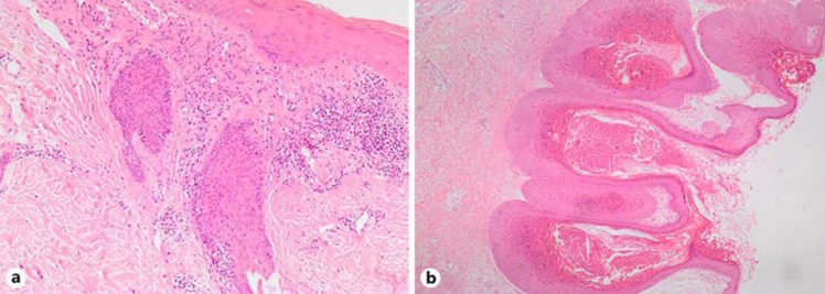
Fig. 2.
Histopathologic images. a HE stain 100×, folliculotropic mycosis fungoides: atypical lymphocytes infiltrating the epithelial layer of the hair follicle. b HE stain 100×, molluscum contagiosum: central umbilication and epidermal hyperplasia. The keratinocytes contain a large intracytoplasmic inclusion compressing the nucleus.

