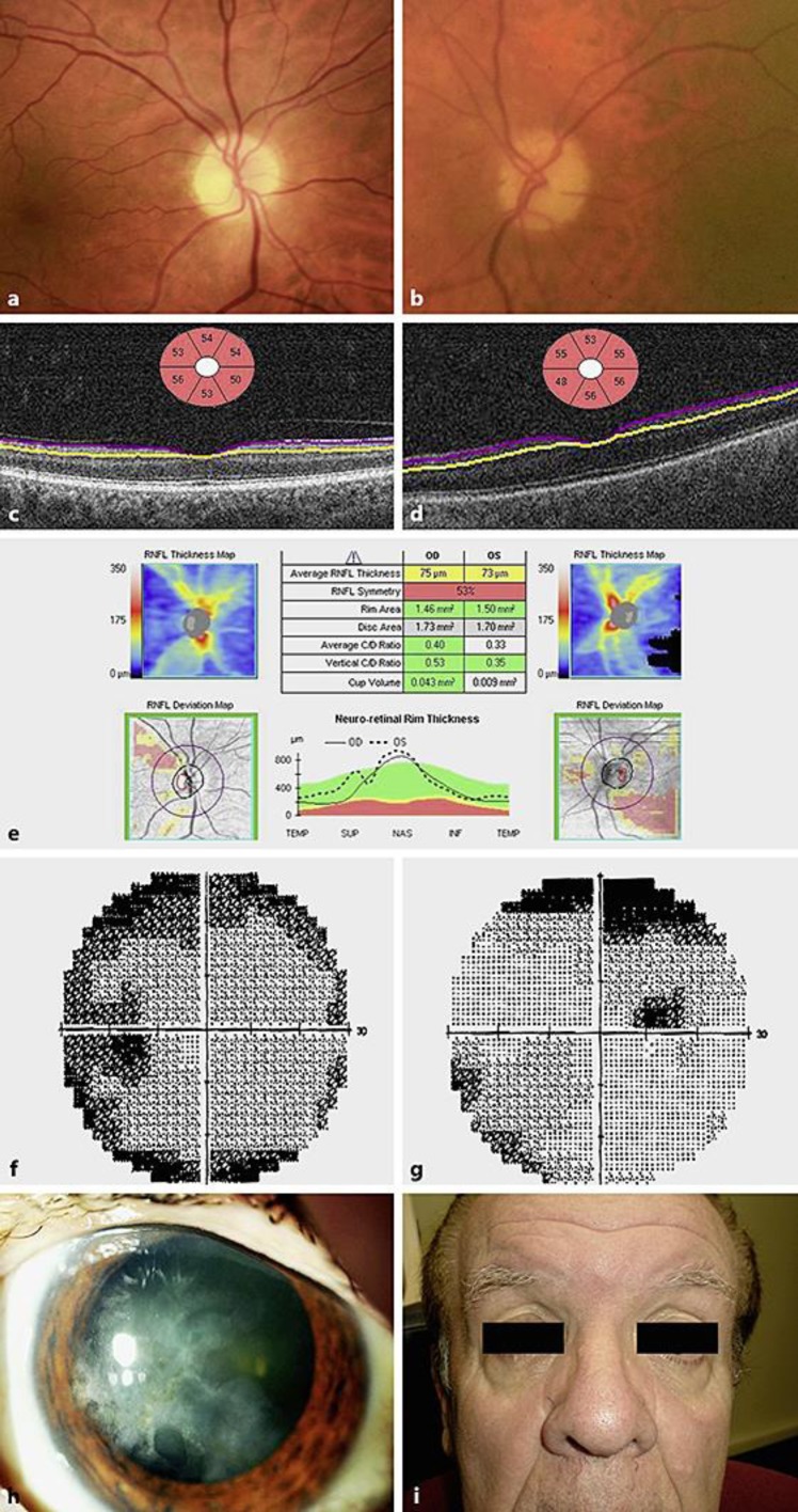
Fig. 1.
A 65-year-old male with bilateral syphilitic optic neuropathy at presentation. Right (a) and left (b) colour fundus photographs showing mild optic disc pallor. Left view is blurred due to old herpetic keratitis. Ganglion cell layer scanning (Cirrus optical coherence tomographic scanning) of the right (c) and left eye (d). e Nerve fibre layer scanning of both eyes showing mild atrophy at presentation. Left (f) and right (g) automated visual field testing (Humphrey) showing a right superior defect. Reliability indices were poor. Colour photograph of the left cornea showing chronic herpes simplex keratitis scarring (h). Facial appearance showing lack of stigmata of congenital syphilis (i).

