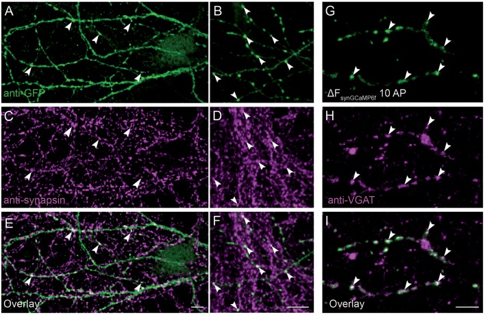
Figure 2.
Presynaptic localization of synGCaMP6f. (A,B) Anti-green fluorescent protein (GFP) antibodies were used to enhance the synGCaMP6f signal (green) and showed cell processes with various varicosities, presumably mostly at presynaptic boutons. (C,D) To verify this, we performed double labeling with anti-synapsin1 (magenta). (E,F) Synaptic GFP-positive varicosities were also immunopositive for synapsin1 (white, arrowheads), indicating the enrichment of synGCaMP6f in presynaptic boutons. (G) The subtraction figure (ΔF, like in Figure 1D) indicates regions with synGCaMP6f fluorescence increase during stimulation with 10 AP. (H) GABAergic presynaptic boutons are identified by incubation with an antibody against VGAT::oyster prior to calcium imaging. (I) The overlay (white) identifies the localization of the synGCaMP6f fluorescence transients (green) to the VGAT-positive GABAergic presynaptic compartments (magenta). Note that no all GABAergic presynapses in this area belong to the neuron that was transfected with synGCaMP6f, as numerous VGAT-positive boutons show no synGCaMP6f signal. Scale bars in (E,F,I): 10 μm.

