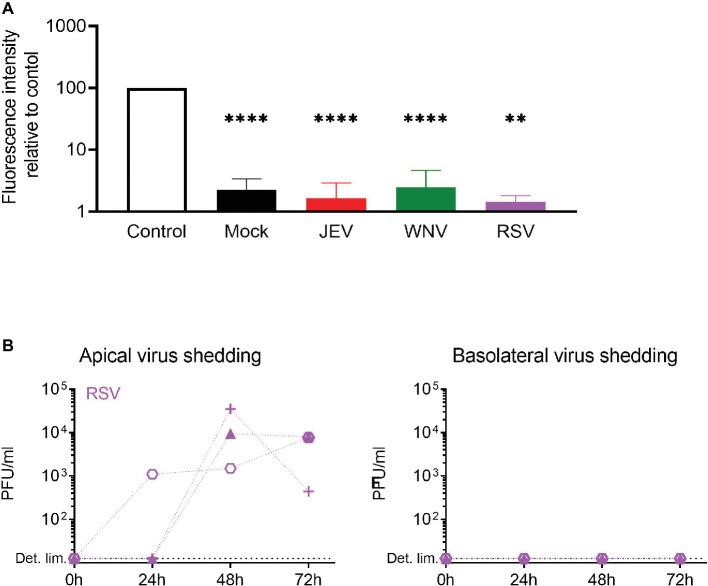
Figure 5.
Epithelial barrier function is maintained upon flavivirus infection. (A) Relative fluorescence intensity in the basolateral chamber was measured after apical application for 4 h of FD4 on inserts 72 h p.i. with JEV, WNV, and RSV. Values are expressed as % of fluorescence relative to control (empty insert). Data are presented as mean and SD of 3–4 healthy donors. (B) Apical and basolateral infectious virus shedding up to 72 h p.i. with RSV measured by PFU assay and expressed in PFU/ml in NECs infected at a MOI of 1 PFU/cell. Each symbol represents an independent donor. Statistical analysis was done in comparison to control; **p < 0.01; ****p < 0.0001.

