Figure 2.
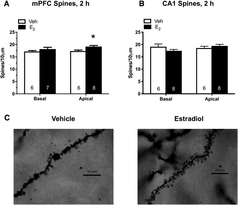
mPFC E2 infusion increases apical spine density in the mPFC 2 h later. Relative to vehicle (Veh), apical but not basal, spine density was significantly increased in the mPFC 2 h after mPFC infusion of 5-µg E2 per hemisphere (A). mPFC infusion did not alter apical or basal spine density in CA1 2 h after mPFC infusion of E2 (B). Bars represent the mean ± SEM; *p < 0.05 relative to the vehicle group (n = 6–8/group). C, Representative photomicrographs of Golgi-impregnated secondary apical dendrites from pyramidal cells in Layer II/III of the mPFC. Under oil 100×.

