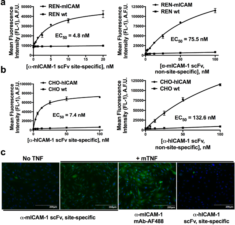
Figure 4.
Affinity and specificity of C-terminal modified scFv–linker–LPETGG. Left panels show flow cytometry based binding curves of sitespecifically modified (a) α-mICAM-1 scFv and (a) α-hICAM-1 scFv to mouse and human ICAM-1 expressing cells, respectively. Binding is specific, with essentially no fluorescent signal seen on wild-type cells. The right panels show NHS-ester modified scFv–linker–LPETGG, which, despite low-level modification (degree of labeling of 1–2 fluorophores per scFv), demonstrates compromised binding affinities. (c) Immunofluorescence of cytokine-activated MS1 mouse endothelial cells shows a similar pattern of staining for site-specifically modified α-mICAM-1 scFv, as compared to fluorescent parental antibody, although the latter is somewhat brighter due to use of a different fluorophore (AlexaFluor 488 vs FITC) and a higher degree of modification. Staining is specific for mouse ICAM-1, with no signal seen for the non-species cross-reactive α-hICAM-1 scFv.

