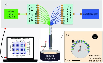
Fig. 2.
(a) Schematic representations of the optical system showing the white-light source and the spectrometer connected to mechanical fiber switches allowing a wide-angle interrogation when the needle is immersed in a tissue phantom composed of multiple inclusions (black carbon rods) embedded in a scattering medium (Intralipid™). (b) Geometrical test configuration around the optical biopsy needle where each of the nine potentially active fiber sources are identified with an even number, whereas the nine detection sources are identified with an odd number. Those numbers are associated with the numbering scheme used for identifying the optical switch positions in (a).

