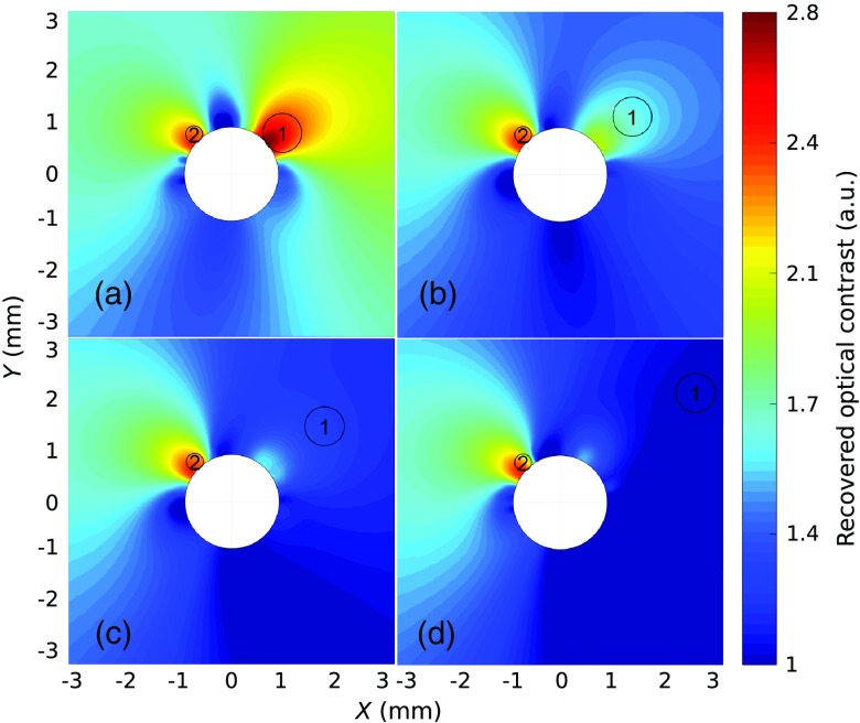
Fig. 3.
Reconstructed diffuse optical images of the carbon rods immersed in a diffusive medium. The distances (edge to edge) between the optical biopsy needle and inclusion 1 are (a) 0 mm, (b) 0.5 mm, (c) 1 mm, and (d) 2 mm. The other carbon rod (inclusion 2) remains at the same position in all experiments.

