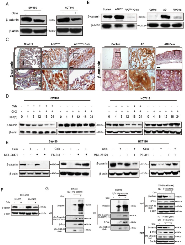
Figure 2.
Celastrol induced β-catenin degradation through the ubiquitin–proteasome pathway.
(A) Western blot of β-catenin in SW480 and HCT116 cells treated with or without Celastrol (0.75 μM) for 24 h. β-actin was used as a loading control.
(B) β-catenin protein levels in intestinal tissues of APCMin/+ mice and colorectal tissues of AOM/DSS mice, with or without Celastrol treatment. β-actin was used as a loading control. N: normal C57 mice; A: APCMin/+ mice; AC: APCMin/+ mice treated with Celastrol (1 mg/kg); AD: AOM/DSS mice; AD + Cela: AOM/DSS mice treated with Celastrol (1 mg/kg).
(C) Immunohistochemistry staining of β-catenin in intestinal tumor tissues of APC Min/+ mice and colon tumor tissues of AOM/DSS mice, treated with or without Celastrol (1 mg/kg), respectively. Wildtype C57 mice were used as a control. AD: AOM/DSS mice. Scale bar: 50 μm (upper), 30 μm (bottom).
(D) The effect of cycloheximide (CHX)(10μg/ml) alone or in combination with Celastrol (0.75 μM) on β-catenin expression were evaluated in SW480 and HCT116 cells at indicated time. β-actin was used as a loading control.
(E) The effect of MDL-28170 (50 μM)/PS-341 (100 nM) alone or in combination with Celastrol (0.75 μM) on β-catenin expression were evaluated in SW480 and HCT116 cells. β-actin was used as a loading control.
(F) HEK293 cells were transfected with widetype or mutant (K48R) ubiquitin plasmids for 24 h, and treated with or without Celastrol (0.75 μM) for another 24 h, respectively. β-catenin were detected and β-actin was used as a loading control.
(G) SW480 and HCT116 cells were treated with or without Celastrol (0.75 μM) for 24 h. Cell lysates were subjected to immunoprecipitation with β-catenin antibody or protein G-Sepharose (IgG as control), followed by immunoblotting with antibodies against ubiquitin, pho-β-catenin (Ser33/37/Thr41), β-Trcp, and pho-GSK-3β (Ser9). Total lysates were used as input control.

