Figure 4.
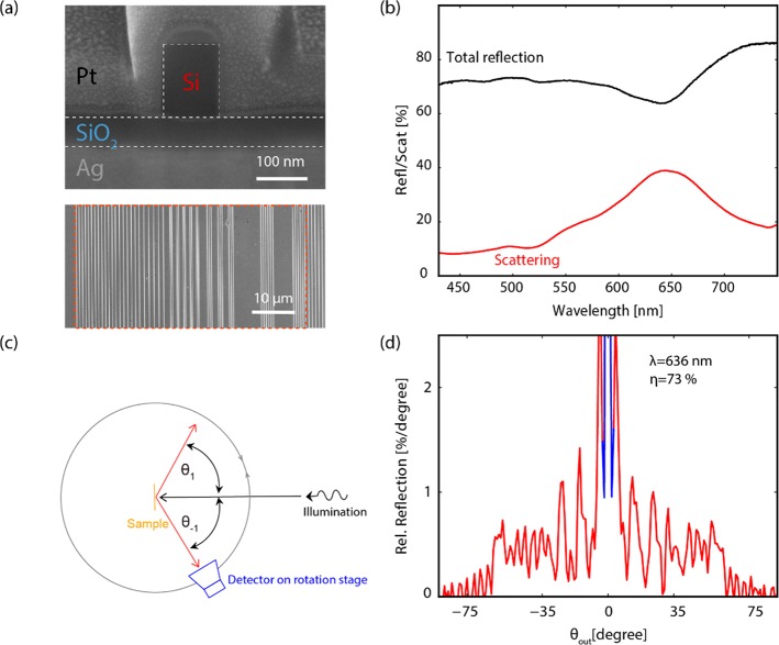
Fabricated broad-angle metasurface and optical measurements. (a) Top: Cross-section SEM image of a silicon bar on a SiOx layer on Ag. To create image contrast. the sample was covered with platinum before performing the FIB cross-section. Bottom: Top-view SEM image. The red dashed box represents a supercell composed of 7 differently pitched metagratings. (b) Total reflection (black) and scattering excluding specular reflection (red). (c) Schematic of angular scattering setup. The sample in the center of a rotating stage is illuminated with a vertical tilt of 10° by a collimated p-polarized light beam (spectral bandwidth 2.5–5 nm). The detector on the rotating stage with a vertical tilt of −10° collects the reflected power at a given horizontal angle θout. (d) Measured angular reflection on resonance (λ = 636 nm). The red data indicate the diffracted scattering, and the blue data are specular reflection.

