Figure 2.
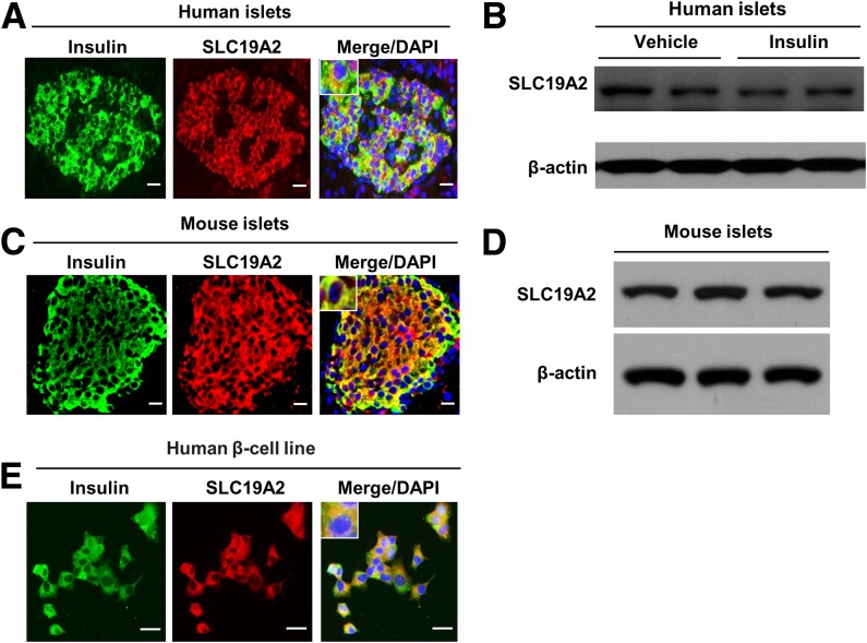
SLC19A2 expression in human and mouse islets. Representative immunostaining for insulin (green), SLC19A2 (red), and nucleus (blue) and Western blot analysis of endocrine pancreas (n = 3). Immunostaining for SLC19A2 and insulin (A) and Western blot analysis of SLC19A2 in human islets (B). Immunostaining for SLC19A2 and insulin (C) and Western blot analysis of SLC19A2 in mouse islets (D). D: Immunostaining for SLC19A2 and insulin in human β-cell line. Scale bars = 20 μm.

