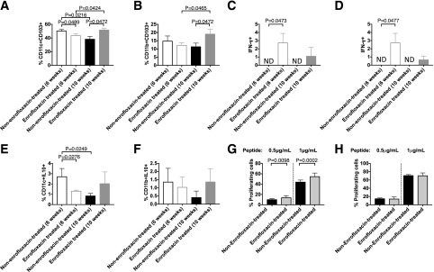
Figure 3.
Direct ex vivo assessment of APC phenotype in untreated and enrofloxacin-treated A22Cα−/−PI2−/− NOD mice. CD11c+ cells or CD11b+ cells were isolated from the spleens of enrofloxacin-treated or untreated A22Cα−/−PI2−/− NOD mice and cocultured in vitro with CFSE-labeled G9CD8+ T cells for 72 h in the presence of insulin B15-23 peptide. A: Frequency of CD11c+CD103+ cells gated from live single CD19−TCRβ−MHCII+CD11b−F480− cells. B: Frequency of CD11b+CD103+ cells gated from live single CD19−TCRβ−MHCII+CD11c−F480+ cells. IFN-γ (C) and IL-10 (E) were assessed in CD11c+ cells gated from live single CD19−TCRβ−MHCII+CD11b−F480− cells. IFN-γ (D) and IL-10 (F) were assessed in CD11b+ cells gated from live single CD19−TCRβ−MHCII+CD11c−F480+ cells. All cells shown in A–F were from PPs. CFSE-labeled G9 CD8+ T cells were cocultured with CD11c+ cells (G) or CD11b+ cells (H) from enrofloxacin-treated or untreated A22Cα−/−PI2−/− NOD mice in a 1:1 ratio in the presence or absence of insulin B15-23 peptide for 48 h. Data shown were corrected for background (cells cocultured without peptide). All statistical analyses were conducted using a Student t test. All data were pooled from two independent experiments (n = 10). Data are presented as mean ± SEM. ND, not detected.

