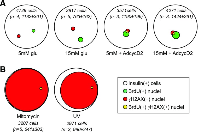
Figure 8.
In human β-cells, proliferation was infrequent even under stimulated conditions, and γH2AX labeling was mostly independent of BrdU-labeled cells. The data in Figs. 6 and 7 are shown using quantitative Venn diagrams to illustrate the relationship between γH2AX labeling and BrdU labeling in these cultures. Diagrams represent the total number (sum of all replicates) of insulin(+) cells (white) that labeled with BrdU (green), γH2AX (red), and both labels (yellow) under different culture conditions. The total number of insulin(+) cells counted, summed for all biological replicates (n = 3–5), is included for each diagram (in italics). A: Although BrdU labeling increased somewhat in proliferative conditions, the γH2AX-labeling index did not, and most γH2AX-labeled cells were not BrdU labeled. B: Mitomycin C and UV irradiation caused DNA damage in the majority of β-cells, but did not increase, in fact decreased, the BrdU-labeling fraction.

