Figure 6.
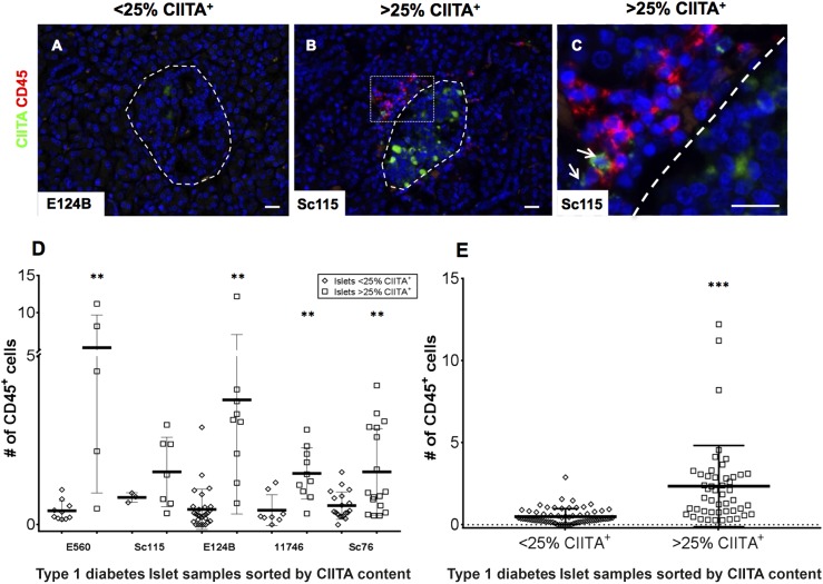
The number of CIITA+ islet cells in sections of pancreas from individuals with type 1 diabetes correlates with the presence of infiltrating immune cells. A–C: Representative images of pancreas sections from five donors with type 1 diabetes stained for CD45 (red) or CIITA (green). Islets are highlighted with dashed lines, and the images depict islets with either <25% of CIITA+ cells (A) or those containing >25% CIITA+ cells (B). C: An enlargement of the highlighted area (dotted white box) is also presented. Arrows indicate cells among the immune cell infiltrate positive for CIITA. Images were captured at ×200 magnification. Multiple <25% and >25% CIITA+ islets were imaged in each of five pancreata from donors with type 1 diabetes and the number of CD45+ cells within (or immediately adjacent to) each islet was counted. Data are shown for each individual case studied (D) and for all cases combined (E). Bars indicate the mean values ±SEM. Scale bars, 20 μm. **P < 0.01, ***P < 0.001.

