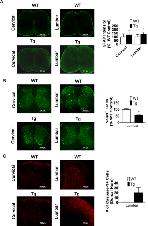
Fig. 9.
Increased GFAP staining and caspase-3+ cells correlates with a reduction in NeuN+ cells in the lumbar spinal cord. a GFAP staining and measurements of staining intensity in the cervical and lumbar spinal cord of 6- week-old WT and MeCP2-Tg mice (n = 4). b NeuN immunostaining and quantification of NeuN+ cells in the lumbar spinal cord of 6-week-old WT and MeCP2-Tg mice (n = 4). Since changes in the number of DAPI-stained cells could contribute to the changes in number of NeuN-positive cells, positive cell counts were normalized to DAPI and expressed as a percentage of wild-type littermate controls. c Capase-3+ cells are increased in the lumbar region of the spinal cord of 6-week-old MeCP2-Tg mice compared to the WT (n = 4)

