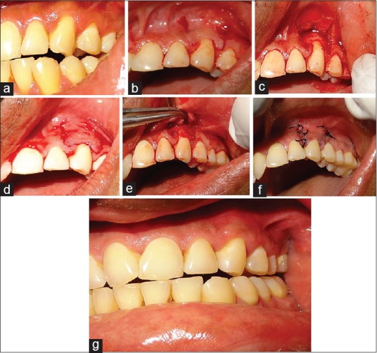
Figure 2.

(a) Photograph showing Miller's Class I gingival recession in 23. (b-d) Operative view showing two beveled vertical releasing incisions on line angle of distal teeth without involving the adjacent papilla extending into the alveolar mucosa. (e) Placement of amnion membrane the root surface. (f) Placement of interrupted sutures to close the vertical incisions. (g) One-year postoperative view showing near complete root coverage in relation to 23 in case 2
