
Figure 5. Inducible expression of SGBP-B* acts in a dominant-negative fashion to inhibit growth in an SGBP-A* strain.
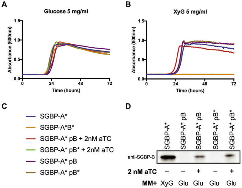
Inducible expression of SGBP-B/B* in an SGBP-A*ΔB background during growth on 5 mg/ml (A) Glucose and (B) XyG. The strains in panel B that did not grow are SGBP-A*/SGBP-B* (orange) and SGBP-A* pSGBP-B* + 2nM aTC (green). (C) Strain legend for panels A,B. (D) Western blot of SGBP-B/B* using anti-SGBP-B serum against whole cell lysates. SGBP-A* strains were cultured in minimal media containing either 5 mg/ml XyG or glucose, with or without 2 nM aTC, as noted. Cells were arrested in mid-logarithmic phase then normalized by O.D.600 before loading equal volumes in SDS-PAGE.