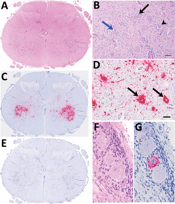
Figure 1.

Histologic and in situ hybridization findings in cervical segment 7 of the spinal cord of 3-week-old lamb in Scotland in study of picornavirus in lambs with severe encephalomyelitis. A) Tranverse segmental view with hematoxylin and eosin stain under low power. B) Hematoxylin and eosin stain under high power. Nonsuppurative myelitis is oriented on the ventral horn involving neuronal degeneration with satellitosis (black arrow), neuronophagia (arrowhead), and glial nodule formation (blue arrow), accompanied by perivascular mononuclear cell accumulation and gliosis. Scale bar indicates 60 μm. C) By in situ hybridization, ovine picornavirus (OvPV) probe hybridization (red) predominates in the region of ventral horn poliomyelitis. D) Viral probe hybridization is dense in neuronal cytoplasm (black arrows) and within presumed neuronal extensions within the neuropil. Scale bar indicates 60 μm. E) Using an unrelated probe, no hybridization is detectable. F, G) Probe hybridization within scattered individual neurons within a spinal ganglion (F, hematoxylin and eosin stain; G, OvPV probe in situ hybridization). Original magnification ×400.
