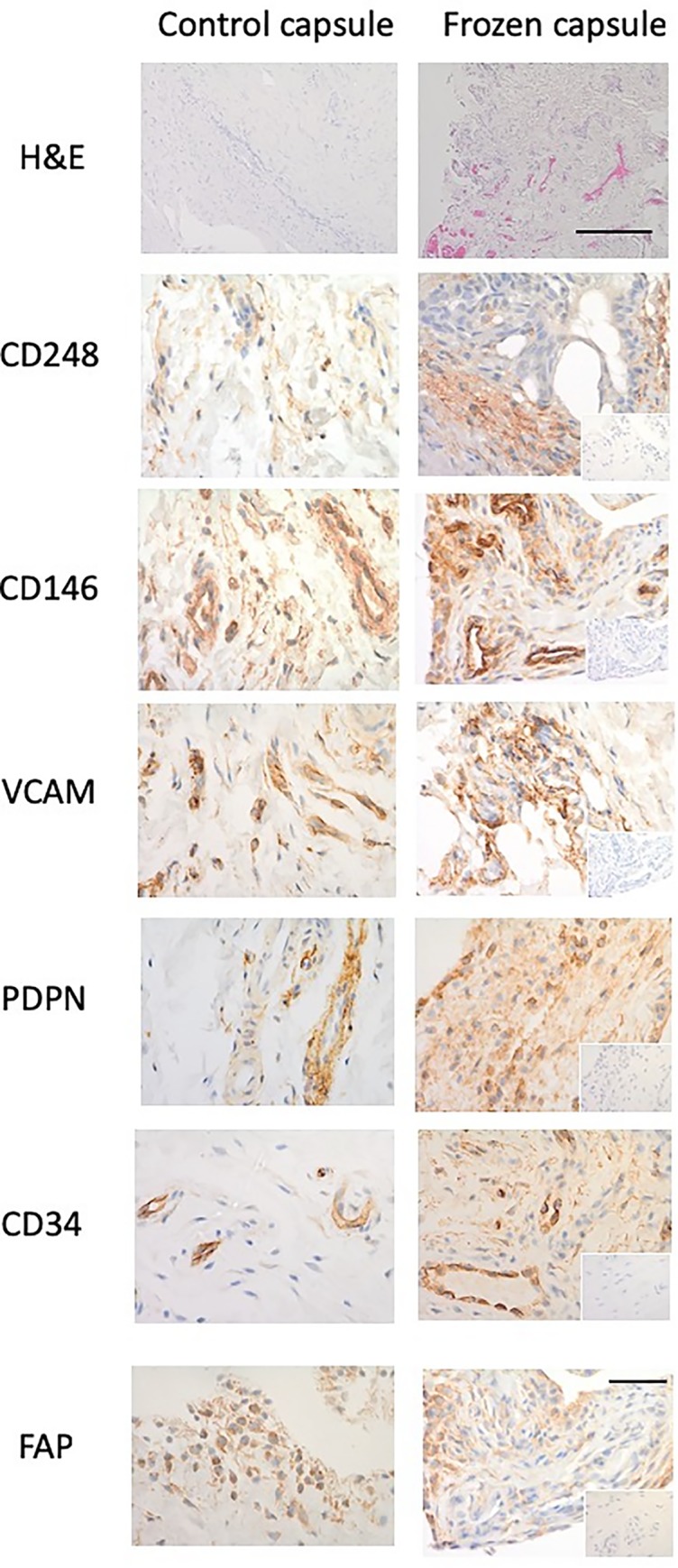
Fig 1. Fibroblast activation markers in shoulder capsule.

Representative images of and control capsule and frozen shoulder tissue sections stained with Haematoxylin and Eosin (x10 magnification, bar indicates 200μm) and antibodies against CD248, CD146, VCAM, PDPN, CD34, and FAP (x40). Isotype control in bottom right corner (x10 magnification, bar indicates 150μm).
