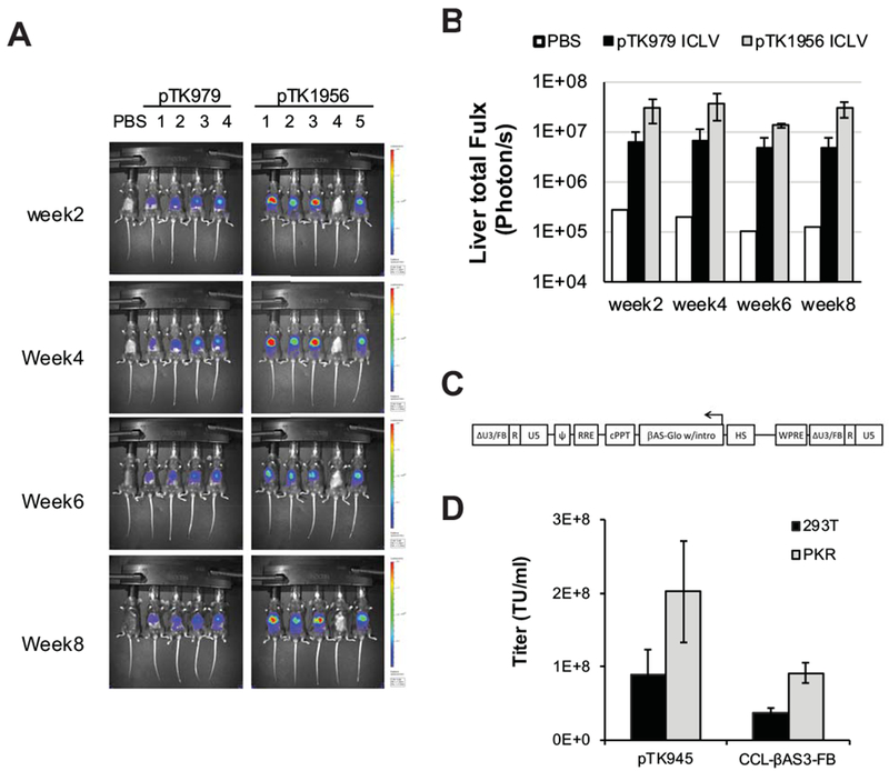
Figure 7. ECOO-comprising lentiviral vectors for hepatic gene delivery and ex-vivo applications.

(A) In vivo imaging of lentiviral vectors-mediated hepatic luciferase expression. C57B6 mice were intraperitoneally injected with 1.5×10⁹ TU of the ECOO-comprising vector pTK1956 (5 mice) and its conventional counterpart pTK979 (4 mice). As shown in figure 5F both vectors carry the firefly luciferase under the control of a liver specific promoter hAAT. A single mouse injected with PBS served as a negative control. Luciferase expression was imaged by the IVIS Lumina optical system at week 2, 4, 6, and 8 post-injection. (B) Bar graph describing average levels of luciferase expression in mouse livers (photon/sec) at weeks 2, 4, 6 and 8 after intraperitoneal administration of lentiviral vectors pTK979 (black bars, average ± SD, n=4) and pTK1956 (grey bars, n=4), and 1 mouse injected with PBS (white bar, n=1). Mouse #4 injected with pTK1956 was excluded from this analysis. (C) Depiction of The CCL-βAS3-FB LV provirus. The corresponding viral vector was developed and described earlier by Romero et al at in the laboratory of Dr. Donald B. Kohn at the University of California, Los Angeles (UCLA). The vector carries the HBBAS3 expression cassette in opposite orientation to the LTR’s (depicted as HBBAS3-Exp). It comprises the β-globin promoter, exons and introns of the β-globin gene as well as its 3’ and 5’ flanking regions including a poly-A site (between the globin coding sequence and the globin 3’ enhancer (Levasseur31 and Behringer 82 et al ). Also included is the β-globin mini-LCR with the hypersensitive sites 2-4. The SIN U3 regions contain a 77 bp mini-insulator (depicted as FB). (D) Bar graph describing titers of VSV-G pseudotyped the ECOO-comprising CCL-βAS3-FB and the conventional vector pTK945 generated in either naïve 293T cells (black bars) or PKR knockdown 293T cells (Grey bars). Error bars represent standard deviation of 2 independent experiments.
