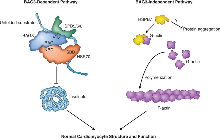
Figure 3. The BAG3-dependent and -independent pathways of cardiac sHSPs.
HSPB5/6/8 represent the BAG3-dependent sHSPs (left), in which HSPB5/6/8 physically and functionally interact with BAG3 to form a multichaperone complex that prevents unfolded substrates from becoming insoluble in cardiomyocytes. HSPB7 represents the BAG3-independent cardiac sHSP (right) that binds to monomeric actin and limits its availability for polymerization. However, it is still unknown whether HSPB7 carries out chaperone activity in vivo. SBD, substrate-binding domain; NBD, nucleotide-binding domain. Illustrated by Rachel Davidowitz.

