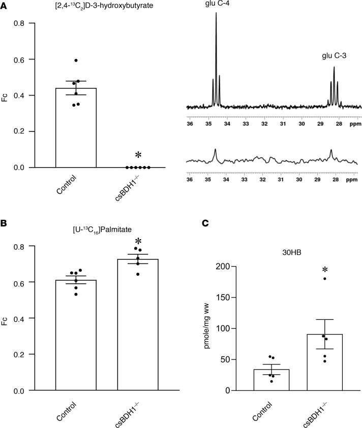
Figure 1. csBDH1–/– mouse hearts are incapable of oxidizing D-3-hydroxybutyrate.
(A) (Left) The fractional enrichment of acetyl-CoA (Fc), representing oxidation of 13C-labeled D-3-hydroxybutyrate into the TCA cycle, is shown in Bdh1fl/flCre– (control) and csBDH1–/– isolated perfused mouse hearts (12- to 16-week-old male littermates) (n = 6). (Right) Representative in vitro NMR spectra displaying 13C labeling of glutamate at the 4- and 3-carbon (glu C-4 and glu C-3) positions in tissue extract from the hearts of control mice (top) and csBDH1–/– mouse (bottom) is shown. The latter has complete absence of signal (1% natural abundance). (B) Fc for 13C-labeled palmitate perfused isolated mouse hearts is shown (n = 5–6) (12- to 16-week-old male littermates). (C) Levels of myocardial 3-hydroxybutyrate (3OHB) per wet weight (ww) measured in control and csBDH1–/– male mice 8–10 weeks after 4-hour fast (n = 5). Bars represent mean ± SEM; *P < 0.05 control vs. csBDH1–/– using unpaired, 2-tailed Mann-Whitney test.

